
Introduction
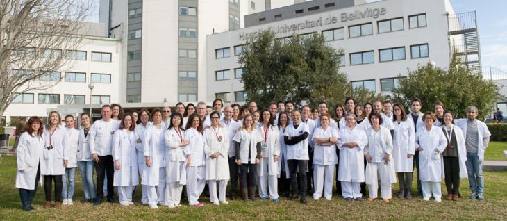
The Ophthalmology Department at the University Hospital of Bellvitge is a Public Service with a high scientific and technological level that provides specialist Ophthalmology healthcare for patients in its area of reference and for all those sent from other centres due to their special complexity, and it combines this service with Education and Research. It has an extensive portfolio of services and top-class technology and equipment to conduct this activity. It also performs research at the IDIBELL Institute, and teaches Medicine and Dentistry Degree studies at the University of Barcelona, as well as training specialist ophthalmology physicians.
Its area of influence for very complex procedures includes a population of reference of 2,242,730 inhabitants. Because the Ophthalmology Department is a centre of reference nationwide (CSUR) for certain ophthalmological diseases, this area of influence also includes other health regions and the rest of Spain.
The HUB Ophthalmology Department aims to become a benchmark healthcare organisation in Catalonia and the rest of Spain, and also worldwide, that stands out for its model of care and its research and educational capacity and its ability to innovate. It strives to ensure this leadership in the field of ophthalmology by developing creativity related to scientific research and innovation in value, as well as by improving specialist training. It also seeks to provide highly specialist ophthalmological care inside and outside the region, helping improve the quality of life of the population and the development of our Region and of the Country.
More information: Barcelona Melanoma Uveal - Translational Research Group in Uveal Melanoma
Related news
Team
Head of Service
Head of Section
Specialist Doctors
Relevant aspects
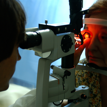
Corneal and anterior segment disorders
Disorders:
- Cataract Keratoconus and other ecstatic disorders, including ecstasia after refractive surgery.
- Fuchs’ endothelial dystrophy.
- Corneal oedema secondary to prior surgery, either a pseudophakic or aphakic bullous keratopathy.
- Hereditary corneal dystrophies.
- Benign or malignant tumours of the ocular surface. Infectious, bacterial, viral or parasitic (Acanthamoeba) keratitis.
- Inflammatory keratitis, including autoimmune pathologies.
- Post-infectious corneal scarring.
- Chemical corneal caustications.
- Corneal neovascularisation.
- Blepharitis and eyelash alterations. Infectious or toxic conjunctivitis
- Acute dry eye disorder (Sjogren’s Sd. arthritis, Graft vs. host disease, etc.)
- Treatment with autologous serum, immunosuppressants such as ciclosporin for acute corneal disorders.
- Corneal pterygium or pseudopterygium
Surgical Techniques:
- Cataract surgery with conventional lens phacoemulsification and
- Femtosecond laser assistance.
- Intraocular lens implant: or Toric lens implant for corneal astigmatism over 3D, o anterior chamber implants in lens subluxations and aphakic patients. Penetrating keratoplasty (PK) in corneal scarring, corneal oedemas, or corneal dystrophies.
- Deep anterior lamellar keratoplasty (DALK) in patients with keratoconus or corneal ecstasies, also in corneal scarring or infectious diseases with a high risk of rejection.
- Endothelial keratoplasty (DSAEK, DMEK) in patients with endothelial diseases such as Fuchs’ dystrophy or bullous keratopathy, or in cases of prior corneal graft rejection.
- Other types of lamellar or penetrating transplantations depending on patient requirements, such as corneal scarring, infectious keratitis, corneal vascularisation, etc.
- Crosslinking treatments in patients with keratoconus or other corneal ecstasies.
- Also possible during post-operative corneal transplantation procedures.
- Resection of benign or malignant corneal or conjunctival tumours. Chemotherapy for the located disease. Conjunctival flaps. Reconstruction of the conjunctival fornix
- Amniotic membrane transplantation (grafts and coverings)
- Corneal limbus transplantation
- Conjunctival autograft transplantation in pterygium surgery
Retina and vitreous
Disorders:
- Diagnosis and treatment of complex cases of age-related macular degeneration (including the variations of proliferative angiomatose retinopathy and polypoidal vasculopathy).
- Diabetic retinopathy and its complications (vitreous haemorrhage, tractional retinal detachment)
- Surgical treatment of complex cases of diabetic retinopathy (tractional retinal detachment, tractional macular oedema, recurrent vitreous haemorrhages) Retinal vein occlusions
- Complex retinal breaks and retinal detachment (giant tears, proliferative vitreoretinopathy, multi-operated patients)
- Complications arising from anterior segment surgery (lens or intraocular lens dislocation towards the vitreous cavity, choroidal detachment, malignant glaucoma, endophthalmitis, expulsive haemorrhage).
- Vitreoretinal interface diseases (epiretinal membrane, macular hole, macular pseudohole, lamellar hole, vitreoretinal traction syndrome). Surgical treatment of complex cases of macular disease (inverted flap, internal limiting membrane translocation, macular indentation)
- High myopia pathologies (Macular hole with or without retinal detachment, myopic retinoschisis and myopic neovascular membrane)
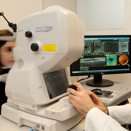
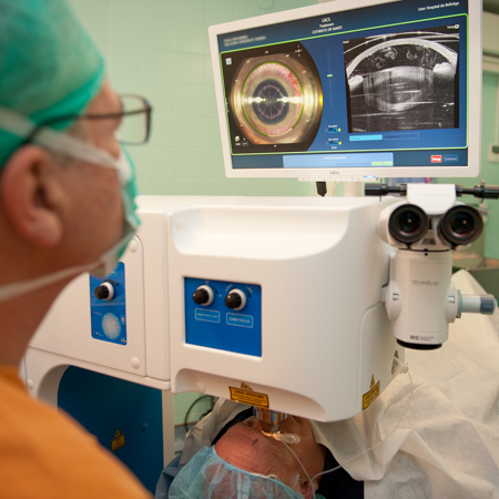
- Retinal dystrophies and degenerative diseases (Conventional electrophysiology and multifocal ERG, angio-OCT and autofluorescence) Intraocular tumours.
- We are a national (CSUR) and European (ERN-EURACAN) centre of reference Intermediate and posterior uveitis.
- We are a benchmark in the biological treatments applied to these patients, as well as in examinations using indocyanine green, swept-source OCT, angio-OCT, and aqueous humor PCR).
- Ocular trauma (intraocular foreign bodies, retinal or choroidal detachment, suprachoroidal haemorrhages)
- Infectious pathology (endophthalmitis)
- Haemorrhagic maculopathies
Surgical Techniques:
- 532 nm green laser
- Photodynamic therapy for treatments of intraocular tumours, serous choroidopathies and choriodal polyps
- Transpupillary thermotherapy for the treatment of intraocular tumours
- Complex 23G and 25G vitrectomy, with or without the application of scleral indentation
- Classic retinal detachment surgery with scleral indentation
- Macular translocation surgery
- Treatment with intraocular antiangiogenic drugs
- Treatment with sustained-release corticosteroid devices Intravitreous, intraretinal and tumour biopsies
- Diagnosis and treatment of complex cases of exudative AMD and macular oedema (angio-OCT, en face, angiography with indocyanine green. Diagnosis of complex cases of retinal dystrophies (ocular electrophysiology, angio-OCT, en face, autofluorescence)
- Surgical treatment of complex causes of retinal detachment (giant tears, PVR, multi-operated patients)
- Surgical treatment of complex cases of diabetic retinopathy (tractional retinal detachment, tractional macular oedema, recurrent vitreous haemorrhages)
- Surgical treatment of complex causes of macular disease (inverted MLI flap, MIL translocation, macular indentation)
- Surgical treatment of complex cases of ocular trauma (intraocular foreign bodies, retinal and choroidal detachment) Sugrical treatment of serious complications from anterior segment surgery (lens and intraocular lens dislocation, endophthalmitis, expulsive haemorrhage, malignant glaucoma)
- We are a benchmark in biological treatments, examinations with indocyanine green and swept-source OCT, angio-OCT and aqueous humor PCR in uveitis.
- Also in difficult-to-diagnose cases as a second opinion. We are a reference in the diagnosis and treatment of intraocular tumours. Especially in opthalmic brachytherapy, and complex tumour resection surgery. (CSUR)
Oculoplastics, lachrymal and orbital
Disorders:
- Orbital tumours
- Thyroid-associated orbitopathy
- Orbital inflammations
- Orbital fractures
- Eyelid malpositions and malfunctions.
- Peripheral facial paralysis (ptosis, retraction, lagophthalmos, blepharospasm, entropion, ectropion)
- Eyelid tumours, especially complex
- Congenital malformations
- Congenital tear duct blockages
- Acquired tear duct blockages
- Tear duct tumours
- Anophthalmic cavity deformities
Surgical Techniques:
- Orbital tumour surgery, including techniques with orbital rim marginotomies, both isolated and extended.
- Microsurgical dissection of orbital tumours.
- Orbital bone and fat decompression.
- In terms of bone decompression, this involve the medial wall, the orbital floor, and the lateral wall.
- Orbital fracture surgery.
- Reduction and reconstruction using autologous and synthetic matter.
- Enophthalmos correction surgery
- Optic nerve sheath fenestration
- Eyelid ptosis surgery Entropion and ectropion surgery
- Entropion and ectropion surgery
- Facial stretching for eyelid reconstruction
- Canthal suspension techniques
- Eyelid laxity techniques and reinsertion of lower eyelid retractors.
- Resection of eyelid tumours and reconstruction of defects
- Symblepharon reconstruction
- Lagophthalmos surgery
- Dacryocystorhinostomy and Dacryocystectomy
- Catheterisation, intubation of the tear duct and balloon dacryocystoplasty
- Canalicular section reconstruction.
- Lester-Jones tubes for upper tear duct occlusions.
- Evisceration, Enucleation and Exenteration
- Reconstruction of anophthalmic cavities with oral mucous grafts
- Reconstructions for the exposition of orbital implants in anophthalmic cavities
Neuro-ophthalmology and ocular motility
Disorders:
This unit sees patients from other centres with undetermined vision loss.
Study of the visual function of pituitary pathology, epilepsy surgery and intracranial tumours
Screening of ocular toxicity in atypical tuberculosis drugs
Visual assessment of patients from the multiple sclerosis and radiotherapy oncology units.
Centre of reference in the treatment of neurovascular pathology (fistulas and aneurysms)
Diagnosis and treatment of complex optical neuropathies (hereditary, inflammatory, paraneoplastic and pseudo-neuropathies)
Complex cases for the treatment of strabismus, nystagmus, and oculomotor nerve palsies.
Study of ocular mitochondrial diseases and other infranuclear disorders
Surgical Techniques:
Horizontal and oblique strabismus surgery
Nystagmus surgery
Oculomotor nerve palsy replacement techniques
Botulinum toxin injections
Strabismus and complex motility disorders (repeat surgeries)
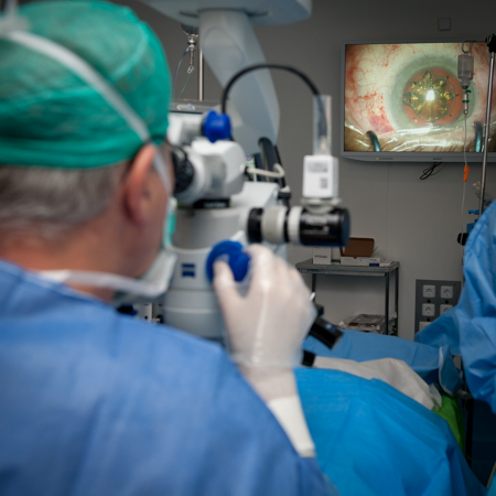
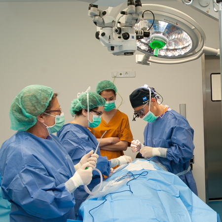
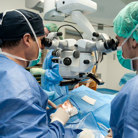
Ophthalmology surgery with femtosecond laser
- A femtosecond laser has been available since March 2018, which enables us to cover cataract surgery with greater safety and efficiency, and means that highly complex corneal surgery is possible with a precision that was unthinkable with manual techniques (especially lamellar corneal transplantation). This device is the first to be installed in a Healthcare Centre of the Public network of Catalonia.
Units of national and European reference
- National CSUR centre of reference in orbital tumours.
- National CSUR centre of reference in orbital decompression.
- National CSUR centre of reference in complex ocular surface pathologies.
- National CSUR centre of reference in in adult-onset intraocular tumours.
- European centre of reference in the treatment of uveal melanoma, with ICO-HUB being a part of the European Reference Network (ERN)- EURACAN Group (rare adult solid cancers).
- Recognised as an Emerging Research Group by the Agency of University Research Grant Management (AGAUR) in the 2017 call.
Multi-disciplinary Units
- Collaboration with the Catalan Oncology Institute (ICO) in the treatment of intraocular tumours.
- Partnership agreement with the San Joan de Déu Hospital in Esplugues de Llobregat to form joint Units for:
- Retinal dystrophies.
- Ocular oncology.
- Complex ocular surface.
- Corneal dystrophies.
- These agreement aim to provide continued healthcare during the transition from childhood to adulthood in these diseases.
- Collaboration with the Blood and Tissue Bank of Catalonia.
- Collaboration with the multiple sclerosis unit.
- Collaboration with the Internal Medicine and Rheumatology Department to treat uveitis.
- Collaboration with the Minority Diseases Unit, with the Retinal dystrophies unit.
- Collaboration with the Multidisciplinary Familial Amyloidosis Unit (MFAU).

Glaucoma
Disorders:
- Diagnosis and treatment of complex primary and secondary glaucomas.
- UBM for the study of the anterior segment.
- Sweep Source-OCT for the study of the pupil, the cribriform plate and angio-OCT.
Surgical Techniques:
- Non-penetrating filtering surgery.
- Trabeculectomy.
- Drainage device implantation.
- Combined cataract-glaucoma surgery.
- Complex glaucoma surgery and surgery for glaucoma complications.
- Amniotic membrane transplantation.
- Microincision surgery.
- Trabeculotomy.
Laser procedures:
- Transscleral cyclophotocoagulation, iridoplasty, trabeculoplasty, suture lysis.
Filtration surgery:
- Experience in all non-penetrating deep sclerectomy techniques, ExPress shunt, trabeculectomy.
- MIGS surgery: XEN stent.
- Drainage devices and transscleral laser cyclophotocoagulation procedures for complex pathologies.
- Endoscopic laser cyclophotocoagulation for complex pathologies.
Research
Lines of research
- Posterior segment section
Basic and translational research focusing into the age-related macular degeneration genome and its relationship with the diagnosis and therapeutic response of anti-VEGF treatment. Basic and translational research into diabetic retinopathy. Degenerative retinal diseases Basic and translational research into the Forms of signalling uveal melanoma and their applicability in establishing new therapies.
- Anterior segment section
Applications of the femtosecond laser in cataract surgery and in corneal pathologies.
This research work primarily involves clinical and basic research work performed at Bellvitge Biomedical Research Institute (IDIBELL), and the implementation of preclinical trials using animal experimentation models, which are conducted in the IDIBELL Animal Housing department.
All this work has led to different presentations and publications in journals of impact.
Main Publications
- Growth of Geographic Atrophy on Fundus Autofluorescence and Polymorphisms of CFH, CFB, C3, FHR1–3, and ARMS2 in Age- Related Macular Degeneration. Spanish Multi-center Group on AMD. JAMA Ophthalmol 2014; 132(5): 528-534.
- Increased fundus autofluorescence and progression of geographic atrophy secondary to age-related macular degeneration. The GAIN study. Biarnés M, Arias L, Alonso J, García M, Hijano M, Rodríguez A, Serrano A, Badal J, Muhtaseb H, Verdaguer P, Monés J. Am J Ophthalmol 2015; 160(2): 345-353.
- International Validation of the American Joint Committee on Cancer's 7th Edition Classification of Uveal Melanoma. The AJCC Ophthalmic Oncology Task Force, Simpson ER, Gallie BL, Saakyan S, Amiryan A, Finger PT, Chin KJ, Seregard S, Fili M, Wilson M, Haik B, Caminal JM, Catala J, Pelayes DE, Folgar MA, Jager M, Dogrusöz M, Singh A, Schachat A, Suzuki S, Aihara Y. JAMA Ophthalmol. 2015; Apr 1;133(4):376-83.
- Local Recurrence Significantly Increases the Risk of Metastatic Uveal Melanoma. Ophthalmic Oncology Task Force. Simpson ER, Gallie BL, Saakyan S, Amiryan A, Finger PT, Chin KJ, Seregard S, Fili M, Wilson M, Haik B, Caminal JM, Catala J, Pelayes DE, Folgar MA, Jager M, Dogrusöz M, Singh A, Schachat A, Suzuki S, Aihara Y.Ophthalmology. 2016 Jan;123(1):86-91
- Tumor volumes in choroidal melanoma: agreement between three-dimensional ultrasound and two mathematical models. Caminal JM, Mejía K, Arias L, Masuet-Aumatell C, Larrucea-Maseda J, Modolell I, Lorenzo D, Català J, Rubio M, Cobos E, García-Bru P, Flores I, Filloy A, Arruga J. Am J Ophthalmol 2016;166:181-188
Education
Docència de Grau
- The Ophthalmology Departmental professors provide education to Medicine and Dentistry Graduates. (Subject: Ophthalmology).
- There is a 4-year Training Programme in the Ophthalmology Speciality: Total 12 Residents. Three Residents per year.
- This is a Resident Rotation Programme that includes Residents from other Hospitals in Catalonia and the rest of Spain.
Master’s degrees and accredited courses
- Master’s degree in Coronary Disease Diagnosis and Interventionist Treatment. Part of the Master’s degree in Advanced Medical Skills at the University of Barcelona
- Specific, accredited fellowship programmes in the following sub-specialities
- Mechanical Ventricular Assistance Training Programmes
- Ongoing training activities: General or specific sessions aimed at medical and nursing staff, residents and fellows International
- Courses organised by the Department
- Fellowship programme: we have accreditation for the post-Residency training of one Retina Fellow under the sponsorship of the Spanish Vitreo-Retinal Society.
Location
Main Building at Bellvitge University Hospital
c/ Feixa Llarga, sn.
08907 L’Hospitalet de Llobregat. Barcelona
Floor 18
Telephone number 93 260 77 05